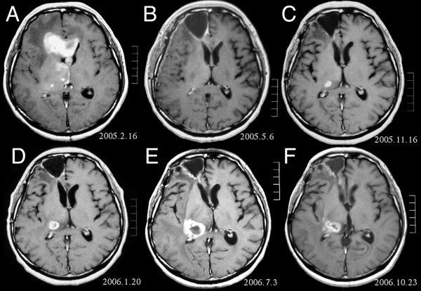
The brain is a soft, spongy mass of tissue. It is protected by the bones of the skull and three thin membranes called meninges. Watery fluid called cerebrospinal fluid cushions the brain. This fluid flows through spaces between the meninges and through spaces within the brain called ventricles.
Illustration shows the skull, spinal cord, brain, meninges, ventricles, the fluid between the meninges, and the fluid in the ventricles.
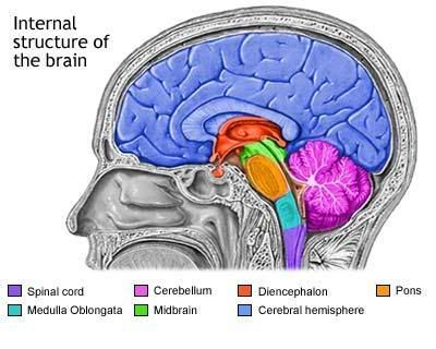
The brain and nearby structures
A network of nerves carries messages back and forth between the brain and the rest of the body. Some nerves go directly from the brain to the eyes, ears, and other parts of the head. Other nerves run through the spinal cord to connect the brain with the other parts of the body. Within the brain and spinal cord, glial cells surround nerve cells and hold them in place.
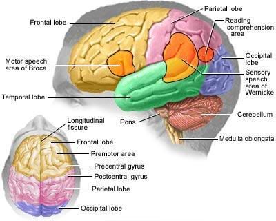
The brain directs the things we choose to do (like walking and talking) and the things our body does without thinking (like breathing). The brain is also in charge of our senses (sight, hearing, touch, taste, and smell), memory, emotions, and personality.
The three major parts of the brain control different activities:
Cerebrum—The cerebrum is the largest part of the brain. It is at the top of the brain. It uses information from our senses to tell us what is going on around us and tells our body how to respond. It controls reading, thinking, learning, speech, and emotions.
 The cerebrum is divided into the left and right cerebral hemispheres, which control separate activities. The right hemisphere controls the muscles on the left side of the body. The left hemisphere controls the muscles on the right side of the body.
Cerebellum—The cerebellum is under the cerebrum at the back of the brain. The cerebellum controls balance and complex actions like walking and talking.
Brain Stem—The brain stem connects the brain with the spinal cord. It controls hunger and thirst. It also controls breathing, body temperature, blood pressure, and other basic body functions.